THE PHOTOSYNTHETIC
PROCESS
In:
"Concepts in Photobiology: Photosynthesis and
Photomorphogenesis", Edited by GS Singhal, G Renger, SK
Sopory, K-D Irrgang and Govindjee, Narosa Publishers/New Delhi;
and Kluwer Academic/Dordrecht, pp. 11-51.
John
Whitmarsh
Photosynthesis Research Unit, Agricultural Research Service/USDA
Department of Plant Biology and Center of Biophysics and
Computational Biology,
University of Illinois at Urbana-Champaign
Govindjee
Department of Plant Biology and Center of Biophysics and
Computational Biology
University of Illinois at Urbana-Champaign
Summary
The primary source of energy for nearly all life is the Sun.
The energy in sunlight is introduced into the biosphere by a
process known as photosynthesis, which occurs in plants, algae
and some types of bacteria. Photosynthesis can be defined as the
physico-chemical process by which photosynthetic organisms use
light energy to drive the synthesis of organic compounds. The
photosynthetic process depends on a set of complex protein
molecules that are located in and around a highly organized
membrane. Through a series of energy transducing reactions, the
photosynthetic machinery transforms light energy into a stable
form that can last for hundreds of millions of years. This
introductory chapter focuses on the structure of the
photosynthetic machinery and the reactions essential for
transforming light energy into chemical energy.
Table of Contents
3.1 Oxygenic Photosynthetic Organisms
3.2 Anoxygenic Photosynthetic Organisms
5.1 Chloroplasts - Structure and Organization
5.2 Light Absorption - The Antenna System
5.3 Primary Photochemistry - Photosystem II and
Photosystem I Reaction Centers
5.4 Electron Transport
5.5 Creation of a Proton Electrochemical
Potential
5.6 Synthesis of ATP by the ATP-Synthase Enzyme
5.7 Synthesis of Carbohydrates
5.8 Photosynthetic Quantum Yields, Energy
Conversion Efficiency and Productivity
5.9 Oxygenic Photosynthesis in Algae
5.10 Oxygenic Photosynthesis in Bacteria
6.1 Purple Bacteria
6.2 Green Sulfur Bacteria
6.3 Green Gliding Bacteria
6.4 Heliobacteria
Photosynthesis is the physico-chemical process by
which plants, algae and photosynthetic bacteria use light energy
to drive the synthesis of organic compounds. In plants, algae and
certain types of bacteria, the photosynthetic process results in
the release of molecular oxygen and the removal of carbon dioxide
from the atmosphere that is used to synthesize carbohydrates
(oxygenic photosynthesis). Other types of bacteria use light
energy to create organic compounds but do not produce oxygen
(anoxygenic photosynthesis). Photosynthesis provides the energy
and reduced carbon required for the survival of virtually all
life on our planet, as well as the molecular oxygen necessary for
the survival of oxygen consuming organisms1 . In addition, the
fossil fuels currently being burned to provide energy for human
activity were produced by ancient photosynthetic organisms.
Although photosynthesis occurs in cells or organelles that are
typically only a few microns across, the process has a profound
impact on the earth's atmosphere and climate. Each year more than
10% of the total atmospheric carbon dioxide is reduced to
carbohydrate by photosynthetic organisms. Most, if not all, of
the reduced carbon is returned to the atmosphere as carbon
dioxide by microbial, plant and animal metabolism, and by biomass
combustion. In turn, the performance of photosynthetic organisms
depends on the earth's atmosphere and climate. Over the next
century, the large increase in the amount of atmospheric carbon
dioxide created by human activity is certain to have a profound
impact on the performance and competition of photosynthetic
organisms. Knowledge of the physico-chemical process of
photosynthesis is essential for understanding the relationship
between living organisms and the atmosphere and the balance of
life on earth. Several books on photosynthesis are available for
the uninitiated (Hall and Rao, 1994; Lawlor, 1993; and Walker,
1992) or advanced student (Govindjee, 1982; Amesz, 1987; Briggs,
1989; Barber, 1992; Scheer, 1991; Bryant, 1994; Blankenship et
al. 1995; Amesz and Hoff, 1996, Baker, 1996; and Ort and Yocum,
1996). Taiz and Zeiger (1991) place the photosynthetic process in
the context of over all plant physiology, and Cramer and Knaff
(1991) describe the bioenergetic foundation of photosynthesis.
The overall equation for photosynthesis is
deceptively simple. In fact, a complex set of physical and
chemical reactions must occur in a coordinated manner for the
synthesis of carbohydrates. To produce a sugar molecule such as
sucrose, plants require nearly 30 distinct proteins that work
within a complicated membrane structure. Research into the
mechanism of photosynthesis centers on understanding the
structure of the photosynthetic components and the molecular
processes that use radiant energy to drive carbohydrate
synthesis. The research involves several disciplines, including
physics, biophysics, chemistry, structural biology, biochemistry,
molecular biology and physiology, and serves as an outstanding
example of the success of multidisciplinary research. As such,
photosynthesis presents a special challenge in understanding
several interrelated molecular processes.
In the 1770s Joseph Priestley, an English chemist
and clergyman, performed experiments showing that plants release
a type of air that allows combustion. He demonstrated this by
burning a candle in a closed vessel until the flame went out. He
placed a sprig of mint in the chamber and after several days
showed that the candle could burn again. Although Priestley did
not know about molecular oxygen, his work showed that plants
release oxygen into the atmosphere. It is noteworthy that over
200 years later, investigating the mechanism by which plants
produce oxygen is one of the most active areas of photosynthetic
research. Building on the work of Priestley, Jan Ingenhousz, a
Dutch physician, demonstrated that sunlight was necessary for
photosynthesis and that only the green parts of plants could
release oxygen. During this period Jean Senebier, a Swiss
botanist and naturalist, discovered that CO2 is required for
photosynthetic growth and Nicolas- Théodore de Saussure, a Swiss
chemist and plant physiologist, showed that water is required. It
was not until 1845 that Julius Robert von Mayer, a German
physician and physicist, proposed that photosynthetic organisms
convert light energy into chemical free energy. An interesting
time line of the history of photosynthesis has been presented by
Huzisige and Ke (1993).
By the middle of the nineteenth
century the key features of plant photosynthesis were known,
namely, that plants could use light energy to make carbohydrates
from CO2 and water. The empirical equation representing the net
reaction of photosynthesis for oxygen evolving organisms is :
CO2 + 2H2O + Light Energy
______> [CH2O] + O2 + H2O, (1)
where [CH2O] represents a
carbohydrate (e.g., glucose, a six-carbon sugar). The synthesis
of carbohydrate from carbon and water requires a large input of
light energy. The standard free energy for the reduction of one
mole of CO2 to the level of glucose is +478 kJ/mol. Because
glucose, a six carbon sugar, is often an intermediate product of
photosynthesis, the net equation of photosynthesis is frequently
written as :
6CO2 + 12H2O + Light Energy
_____> C6H12O6 + 6O2 + 6H2O. (2)
The standard free energy for the synthesis of
glucose is +2,870 kJ/mol.
Not surprisingly, early scientists
studying photosynthesis concluded that the O2 released by plants
came from CO2, which was thought to be split by light energy. In
the 1930s comparison of bacterial and plant photosynthesis lead
Cornelis van Niel to propose the general equation of
photosynthesis that applies to plants, algae and photosynthetic
bacteria (discussed by Wraight, 1982). Van Niel was aware that
some photosynthetic bacteria could use hydrogen sulfide (H2S)
instead of water for photosynthesis and that these organisms
released sulfur instead of oxygen. Van Niel, among others,
concluded that photosynthesis depends on electron donation and
acceptor reactions and that the O2 released during photosynthesis
comes from the oxidation of water. Van Niel's generalized
equation is :
CO2 + 2H2A + Light Energy
_____> [CH2O] + 2A + H2O. (3)
In oxygenic photosynthesis, 2A is O2, whereas in
anoxygenic photosynthesis, which occurs in some photosynthetic
bacteria, the electron donor can be an inorganic hydrogen donor,
such as H2S (in which case A is elemental sulfur) or an organic
hydrogen donor such as succinate (in which case, A is fumarate).
Experimental evidence that molecular oxygen came from water was
provided by Hill and Scarisbrick (1940) who demonstrated oxygen
evolution in the absence of CO2 in illuminated chloroplasts and
by Ruben et al. (1941) who used 18O enriched water.
The biochemical conversion of CO2 to carbohydrate
is a reduction reaction that involves the rearrangement of
covalent bonds between carbon, hydrogen and oxygen. The energy
for the reduction of carbon is provided by energy rich molecules
that are produced by the light driven electron transfer
reactions. Carbon reduction can occur in the dark and involves a
series of biochemical reactions that were elucidated by Melvin
Calvin, Andrew Benson and James Bassham in the late 1940s and
1950s. Using the radioisotope 14C, most of the intermediate steps
that result in the production of carbohydrate were identified.
Calvin was awarded the Nobel Prize for Chemistry in 1961 for this
work (see Calvin, 1989).
In 1954 Daniel Arnon and coworkers discovered
that plants, and A. Frenkel discovered that photosynthetic
bacteria, use light energy to produce ATP, an organic molecule
that serves as an energy source for many biochemical reactions
(discussed by Frenkel, 1995). During the same period L.N.M.
Duysens showed that the primary photochemical reaction of
photosynthesis is an oxidation/reduction reaction that occurs in
a protein complex (the reaction center). Over the next few years
the work of several groups, including those of Robert Emerson,
Bessel Kok, L.N.M. Duysens, Robert Hill and Horst Witt, combined
to prove that plants, algae and cyanobacteria require two
reaction centers, photosystem II and photosystem I, operating in
series (Duysens, 1989; Witt, 1991).
In 1961 Peter Mitchell suggested that cells can
store energy by creating an electric field or a proton gradient
across a membrane. Mitchell's proposal that energy is stored as
an electrochemical gradient across a vesicular membrane opened
the door for understanding energy transformation by membrane
systems. He was awarded the Nobel Prize in Chemistry in 1978 for
his theory of chemiosmotic energy transduction (Mitchell, 1961).
Most of the proteins required for the conversion
of light energy and electron transfer reactions of photosynthesis
are located in membranes. Despite decades of work, efforts to
determine the structure of membrane bound proteins had little
success. This changed in the 1980s when Johann Deisenhofer,
Hartmut Michel, Robert Huber and co-workers determined the
structure of the reaction center of the purple bacterium
Rhodospeudomonas viridis. (Deisenhofer et al., 1984, 1985;
Deisenhofer and Michel, 1993). They were awarded the Nobel Prize
for Chemistry in 1988 for their work, which has provided insight
into the relationship between structure and function in
membrane-bound proteins .
A key element in photosynthetic energy conversion
is electron transfer within and between protein complexes and
simple organic molecules. The electron transfer reactions are
rapid (as fast as a few picoseconds) and highly specific. Much of
our current understanding of the physical principles that guide
electron transfer is based on the pioneering work of Rudolph A.
Marcus (Marcus and Sutin, 1985), who received the Nobel Prize in
Chemistry in 1992 for his contributions to the theory of electron
transfer reaction in chemical systems.
All life can be divided into three domains,
Archaea, Bacteria and Eucarya, which originated from a common
ancestor (Woese et al., 1990). Historically, the term
photosynthesis has been applied to organisms that depend on
chlorophyll (or bacteriochlorophyll) for the conversion of light
energy into chemical free energy (Gest , 1993). These include
organisms in the domains Bacteria (photosynthetic bacteria) and
Eucarya (algae and higher plants). The most primitive domain,
Archaea, includes organisms known as halobacteria, that convert
light energy into chemical free energy. However, the mechanism by
which halobacteria convert light is fundamentally different from
that of higher organisms because there is no oxidation/reduction
chemistry and halobacteria cannot use CO2 as their carbon source.
Consequently some biologists do not consider halobacteria as
photosynthetic (Gest 1993). This chapter will follow the
historical definition of photosynthesis and omit halobacteria.
3.1 Oxygenic Photosynthetic
Organisms
The photosynthetic process in all plants and algae as well
as in certain types of photosynthetic bacteria involves the
reduction of CO2 to carbohydrate and removal of electrons from
H20, which results in the release of O2. In this process, known
as oxygenic photosynthesis, water is oxidized by the photosystem
II reaction center, a multisubunit protein located in the
photosynthetic membrane. Years of research have shown that the
structure and function of photosystem II is similar in plants,
algae and certain bacteria, so that knowledge gained in one
species can be applied to others. This homology is a common
feature of proteins that perform the same reaction in different
species. This homology at the molecular level is important
because there are estimated to be 300,000-500,000 species of
plants. If different species had evolved diverse mechanisms for
oxidizing water, research aimed at a general understanding of
photosynthetic water oxidation would be hopeless.
3.2 Anoxygenic
Photosynthetic Organisms
Some photosynthetic bacteria can use light energy to
extract electrons from molecules other than water. These
organisms are of ancient origin, presumed to have evolved before
oxygenic photosynthetic organisms. Anoxygenic photosynthetic
organisms occur in the domain Bacteria and have representatives
in four phyla - Purple Bacteria, Green Sulfur Bacteria, Green
Gliding Bacteria, and Gram Positive Bacteria.
The energy that drives photosynthesis originates
in the center of the sun, where mass is converted to heat by the
fusion of hydrogen. Over time, the heat energy reaches the sun's
surface, where some of it is converted to light by black body
radiation that reaches the earth. A small fraction of the visible
light incident on the earth is absorbed by plants. Through a
series of energy transducing reactions, photosynthetic organisms
are able to transform light energy into chemical free energy in a
stable form that can last for hundreds of millions of years
(e.g., fossil fuels). A simplified scheme describing how energy
is transformed in the photosynthetic process is presented in this
section. The focus is on the structural and functional features
essential for the energy transforming reactions. For clarity,
mechanistic and structural details are omitted. A more highly
resolved description of oxygenic and anoxygenic photosynthesis is
given in the remaining sections.
The photosynthetic process in plants and algae
occurs in small organelles known as chloroplasts that are located
inside cells. The more primitive photosynthetic organisms, for
example oxygenic cyanobacteria, prochlorophytes and anoxygenic
photosynthetic bacteria, lack organelles. The photosynthetic
reactions are traditionally divided into two stages - the
"light reactions," which consist of electron and proton
transfer reactions and the "dark reactions," which
consist of the biosynthesis of carbohydrates from CO2. The light
reactions occur in a complex membrane system (the photosynthetic
membrane) that is made up of protein complexes, electron
carriers, and lipid molecules. The photosynthetic membrane is
surrounded by water and can be thought of as a two-dimensional
surface that defines a closed space, with an inner and outer
water phase. A molecule or ion must pass through the
photosynthetic membrane to go from the inner space to the outer
space. The protein complexes embedded in the photosynthetic
membrane have a unique orientation with respect to the inner and
outer phase. The asymmetrical arrangement of the protein
complexes allows some of the energy released during electron
transport to create an electrochemical gradient of protons across
the photosynthetic membrane.
Photosynthetic electron transport consists of a
series of individual electron transfer steps from one electron
carrier to another. The electron carriers are metal ion complexes
and aromatic groups. The metal ion complexes and most of the
aromatic groups are bound within proteins. Most of the proteins
involved in photosynthetic electron transport are composed of
numerous polypeptide chains that lace through the membrane,
providing a scaffolding for metal ions and aromatic groups. An
electron enters a protein complex at a specific site, is
transferred within the protein from one carrier to another, and
exits the protein at a different site. The protein controls the
pathway of electrons between the carriers by determining the
location and environment of the metal ion complexes and aromatic
groups. By setting the distance between electron carriers and
controlling the electronic environment surrounding a metal ion
complex or aromatic group, the protein controls pairwise electron
transfer reactions. Between proteins, electron transfer is
controlled by distance and free energy, as for intraprotein
transfer, and by the probability that the two proteins are in
close contact. Protein association is controlled by a number of
factors, including the structure of the two proteins, their
surface electrical and chemical properties and the probability
that they collide with one another. Not all electron carriers are
bound to proteins. The reduced forms of plastoquinone or
ubiquinone and nicotinamide adenine dinucleotide phosphate
(NADPH) or NADH act as mobile electron carriers operating between
protein complexes. For electron transfer to occur, these small
molecules must bind to special pockets in the proteins known as
binding sites. The binding sites are highly specific and are a
critical factor in controlling the rate and pathway of electron
transfer.
The light reactions convert energy into several
forms (Fig. 1). The first step is the
conversion of a photon to an excited electronic state of an
antenna pigment molecule located in the antenna system. The
antenna system consists of hundreds of pigment molecules (mainly
chlorophyll or bacteriochlorophyll and carotenoids) that are
anchored to proteins within the photosynthetic membrane and serve
a specialized protein complex known as a reaction center. The
electronic excited state is transferred over the antenna
molecules as an exciton. Some excitons are converted back into
photons and emitted as fluorescence, some are converted to heat,
and some are trapped by a reaction center protein. (For a
discussion of the use of fluorescence as a probe of
photosynthesis, see e.g., Govindjee et al., 1986 and Krause and
Weis, 1991.) Excitons trapped by a reaction center provide the
energy for the primary photochemical reaction of photosynthesis -
the transfer of an electron from a donor molecule to an acceptor
molecule. Both the donor and acceptor molecules are attached to
the reaction center protein complex. Once primary charge
separation occurs, the subsequent electron transfer reactions are
energetically downhill.
In oxygenic photosynthetic organisms (see section 5), two
different reaction centers, known as photosystem II and
photosystem I, work concurrently but in series. In the light
photosystem II feeds electrons to photosystem I. The electrons
are transferred from photosystem II to the photosystem I by
intermediate carriers. The net reaction is the transfer of
electrons from a water molecule to NADP+, producing the reduced
form, NADPH. In the photosynthetic process, much of the energy
initially provided by light energy is stored as redox free energy
(a form of chemical free energy) in NADPH, to be used later in
the reduction of carbon. In addition, the electron transfer
reactions concentrate protons inside the membrane vesicle and
create an electric field across the photosynthetic membrane. In
this process the electron transfer reactions convert redox free
energy into an electrochemical potential of protons. The energy
stored in the proton electrochemical potential is used by a
membrane bound protein complex (ATP-Synthase) to covalently
attach a phosphate group to adenosine diphosphate (ADP), forming
adenosine triphosphate (ATP). Protons pass through the
ATP-Synthase protein complex that transforms electrochemical free
energy into a type of chemical free energy known as phosphate
group-transfer potential (or a high-energy phosphate bond)
(Klotz, 1967). The energy stored in ATP can be transferred to
another molecule by transferring the phosphate group. The net
effect of the light reactions is to convert radiant energy into
redox free energy in the form of NADPH and phosphate
group-transfer energy in the form of ATP. In the light reactions,
the transfer of a single electron from water to NADP+ involves
about 30 metal ions and 7 aromatic groups. The metal ions include
19 Fe, 5 Mg, 4 Mn, and 1 Cu. The aromatics include quinones,
pheophytin, NADPH, tyrosine and a flavoprotein. The NADPH and ATP
formed by the light reactions provide the energy for the dark
reactions of photosynthesis, known as the Calvin cycle or the
photosynthetic carbon reduction cycle. The reduction of
atmospheric CO2 to carbohydrate occurs in the aqueous phase of
the chloroplast and involves a series of enzymatic reactions. The
first step is catalyzed by the protein Rubisco (D-ribulose
1,5-bisphosphate carboxylase/oxygenase), which attaches CO2 to a
five-carbon compound. The reaction produces two molecules of a
three-carbon compound. Subsequent biochemical reactions involve
several enzymes that reduce carbon by hydrogen transfer and
rearrange the carbon compounds to synthesize carbohydrates. The
carbon reduction cycle involves the transfer and rearrangement of
chemical bond energy.
In anoxygenic photosynthetic organisms (see section 6) water
is not used as the electron donor. Electron flow is cyclic and is
driven by a single photosystem, producing a proton
electrochemical gradient that is used to provide energy for the
reduction of NAD+ by an external H-atom or e-donor (e.g., H2S or
an organic acid) in a process known as "reverse electron
flow". Fixation of CO2 occurs via different pathways in
different organisms.
5.1 Chloroplasts -
Structure and Organization
In plants the photosynthetic process occurs inside
chloroplasts, which are organelles found in certain cells.
Chloroplasts provide the energy and reduced carbon needed for
plant growth and development, while the plant provides the
chloroplast with CO2, water, nitrogen, organic molecules and
minerals necessary for the chloroplast biogenesis. Most
chloroplasts are located in specialized leaf cells, which often
contain 50 or more chloroplasts per cell. Each chloroplast is
defined by an inner and an outer envelope membrane and is shaped
like a meniscus convex lens that is 5-10 microns in diameter (Fig. 2), although many different shapes and
sizes can be found in plants. For details of chloroplast
structure, see Staehlin (1986). The inner envelope membrane acts
as a barrier, controlling the flux of organic and charged
molecules in and out of the chloroplast. Water passes freely
through the envelope membranes, as do other small neutral
molecules like CO2 and O2. There is evidence that chloroplasts
were once free living bacteria that invaded a non-photosynthetic
cell long ago. They have retained some of the DNA necessary for
their assembly, but much of the DNA necessary for their
biosynthesis is located in the cell nucleus. This enables a cell
to control the biosynthesis of chloroplasts within its domain.
Inside the chloroplast is a complicated membrane system, known
as the photosynthetic membrane (or thylakoid membrane), that
contains most of the proteins required for the light reactions.
The proteins required for the fixation and reduction of CO2 are
located outside the photosynthetic membrane in the surrounding
aqueous phase. The photosynthetic membrane is composed mainly of
glycerol lipids and protein. The glycerol lipids are a family of
molecules characterized by a polar head group that is hydrophilic
and two fatty acid side chains that are hydrophobic. In
membranes, the lipid molecules arrange themselves in a bilayer,
with the polar head toward the water phase and the fatty acid
chains aligned inside the membrane forming a hydrophobic core (Fig. 3). The photosynthetic membrane is
vesicular, defining a closed space with an outer water space
(stromal phase) and an inner water space (lumen). The
organization of the photosynthetic membrane can be described as
groups of stacked membranes (like stacks of pita or chapati bread
with the inner pocket representing the inner aqueous space),
interconnected by non-stacked membranes that protrude from the
edges of the stacks (Fig. 2). Experiments
indicate that the inner aqueous space of the photosynthetic
membrane is likely continuous inside of the chloroplast. It is
not known why the photosynthetic membrane forms such a convoluted
structure. To understand the energetics of photosynthesis the
complicated structure can be ignored and the photosynthetic
membrane can be viewed as a simple vesicle.
5.2 Light Absorption - The
Antenna System
Plant photosynthesis is driven primarily by visible light
(wavelengths from 400 to 700 nm) that is absorbed by pigment
molecules (mainly chlorophyll a and b and carotenoids). The
chemical structure of chlorophyll a molecule is shown in Fig. 4. In chlorophyll b, CH3 in ring II is
replaced by CHO group. Plants appear green because of
chlorophyll, which is so plentiful that regions of the earth
appear green from space. The absorption spectrum of chloroplast
chlorophyll a and b and carotenoids along with the action
spectrum of photosynthesis of a chloroplast is shown in Fig. 5. Light is collected by 200-300 pigment
molecules that are bound to light- harvesting protein complexes
located in the photosynthetic membrane. The light-harvesting
complexes surround the reaction centers that serve as an antenna.
The three-dimensional structure of the light-harvesting complex
(Kühlbrandt et al., 1994) shows that the protein determines the
position and orientation of the antenna pigments. Photosynthesis
is initiated by the absorption of a photon by an antenna
molecule, which occurs in about a femtosecond (10-15 s) and
causes a transition from the electronic ground state to an
excited state. Within 10-13 s the excited state decays by
vibrational relaxation to the first excited singlet state. The
fate of the excited state energy is guided by the structure of
the protein. Because of the proximity of other antenna molecules
with the same or similar energy states, the excited state energy
has a high probability of being transferred by resonance energy
transfer to a near neighbor. Exciton energy transfer between
antenna molecules is due to the interaction of the transition
dipole moment of the molecules. The probability of transfer is
dependent on the distance between the transition dipoles of the
donor and acceptor molecules (1/R6), the relative orientation of
the transition dipoles, and the overlap of the emission spectrum
of the donor molecule with the absorption spectrum of the
acceptor molecule (see van Grondelle and Amesz, 1986).
Photosynthetic antenna systems are very efficient at this
transfer process. Under optimum conditions over 90% of the
absorbed quanta are transferred within a few hundred picoseconds
from the antenna system to the reaction center which acts as a
trap for the exciton. A simple model of the antenna and its
reaction center is shown in Fig. 6.
5.3 Primary Photochemistry
- Photosystem II and Photosystem I Reaction Centers
Photosystem II uses light energy to drive two chemical
reactions - the oxidation of water and the reduction of
plastoquinone. The photosystem II complex is composed of more
than fifteen polypeptides and at least nine different redox
components (chlorophyll, pheophytin, plastoquinone, tyrosine, Mn,
Fe, cytochrome b559, carotenoid and histidine) have been shown to
undergo light-induced electron transfer (Debus, 1992). However,
only five of these redox components are known to be involved in
transferring electrons from H2O to the plastoquinone pool - the
water oxidizing manganese cluster (Mn)4, the amino acid tyrosine,
the reaction center chlorophyll (P680), pheophytin, and the
plastoquinone molecules, QA and QB. Of these essential redox
components, tyrosine, P680, pheophytin, QA and QB have been shown
to be bound to two key polypeptides that form the heterodimeric
reaction center core of photosystem II (D1 and D2). Recent work
indicates that the D1 and D2 polypeptides also provide ligands
for the (Mn)4 cluster. The three-dimensional structure of
photosystem II is not known. Our knowledge of its structure is
guided by the known structure of the reaction center in purple
bacteria and biochemical and spectroscopic data. Fig. 7 shows a schematic view of photosystem
II that is consistent with current data.
Photochemistry in photosystem II is initiated by charge
separation between P680 and pheophytin, creating P680+/Pheo-.
Primary charge separation takes about a few picoseconds (Fig. 8). Subsequent electron transfer steps
have been designed through evolution to prevent the primary
charge separation from recombining. This is accomplished by
transferring the electron within 200 picoseconds from pheophytin
to a plastoquinone molecule (QA) that is permanently bound to
photosystem II. Although plastoquinone normally acts as a
two-electron acceptor, it works as a one-electron acceptor at the
QA-site. The electron on QA- is then transferred to another
plastoquinone molecule that is loosely bound at the QB-site.
Plastoquinone at the QB-site differs from QA in that it works as
a two-electron acceptor, becoming fully reduced and protonated
after two photochemical turnovers of the reaction center. The
full reduction of plastoquinone requires the addition of two
electrons and two protons, i.e., the addition of two hydrogen
atoms. The reduced plastoquinone (Fig. 9)
then debinds from the reaction center and diffuses into the
hydrophobic core of the membrane. After which, an oxidized
plastoquinone molecule finds its way to the QB-binding site and
the process is repeated. Because the QB-site is near the outer
aqueous phase, the protons added to plastoquinone during its
reduction are taken from the outside of the membrane.
Photosystem II is the only known protein complex that can
oxidize water, resulting in the release of O2 into the
atmosphere. Despite years of research, little is known about the
molecular events that lead to water oxidation. Energetically,
water is a poor electron donor. The oxidation- reduction midpoint
potential (Em,7) of water is +0.82 V (pH 7). In photosystem II
this reaction is driven by the oxidized reaction center, P680+
(the midpoint potential of P680/P680+ is estimated to be +1.2 V
at pH 7). How electrons are transferred from water to P680+
remains a mystery (Govindjee and Coleman, 1990). It is known that
P680+ oxidizes a tyrosine on the D1 protein and that Mn plays a
key role in water oxidation. Four Mn ions are present in the
water oxidizing complex. X-ray absorption spectroscopy shows that
Mn undergoes light-induced oxidation. Water oxidation requires
two molecules of water and involves four sequential turnovers of
the reaction center. This was shown by an experiment
demonstrating that oxygen release by photosystem II occurs with a
four flash dependence (Fig. 10; Joliot et
al., 1969; Joliot and Kok, 1975). Each photochemical reaction
creates an oxidant that removes one electron. The net reaction
results in the release of one O2 molecule, the deposition of four
protons into the inner water phase, and the transfer of four
electrons to the QB-site (producing two reduced plastoquinone
molecules) (reviewed by Renger, 1993; Klein et al., 1993; and
Lavergne and Junge , 1993).
Photosystem II reaction centers contain a number of redox
components with no known function. An example is cytochrome b559,
a heme protein, that is an essential component of all photosystem
II reaction centers (discussed by Whitmarsh and Pakrasi, 1996).
If the cytochrome is not present in the membrane, a stable PS II
reaction center cannot be formed. Although the structure and
function of Cyt b559 remain to be discovered, it is known that
the cytochrome is not involved in the primary enzymatic activity
of PS II, which is the transfer of electrons from water to
plastoquinone. Why PS II reaction centers contain redox
components that are not involved in the primary enzymatic
reactions is a puzzling question. The answer may be found in the
unusual chemical reactions occurring in PS II and the fact that
the reaction center operates at a very high power level.
Photosystem II is an energy transforming enzyme that must switch
between various high energy states that involve the creation of
the powerful oxidants required for removing electrons from water
and the complex chemistry of plastoquinone reduction which is
strongly influenced by protons. In saturating light a single
reaction center can have an energy throughput of 600 eV/s
(equivalent to 60,000 kW per mole of PS II). Operating at such a
high power level results in damage to the reaction center. It may
be that some of the "extra" redox components in
photosystem II may serve to protect the reaction center.
Photosystem II has another perplexing feature. Many plants and
algae have been shown to have a significant number of photosystem
II reaction centers that do not contribute to photosynthetic
electron transport (e.g., Chylla and Whitmarsh, 1989). Why plants
devote resources for the synthesis of reaction centers that
apparently do not contribute to energy conversion is unknown (for
reviews of photosystem II heterogeneity see Ort and Whitmarsh,
1990; Guenther and Melis, 1990; Govindjee, 1990; Melis, 1991;
Whitmarsh et al., 1996; Lavergne and Briantais, 1996)
The photosystem I complex catalyzes the oxidation of
plastocyanin, a small soluble Cu- protein, and the reduction of
ferredoxin, a small FeS protein (Fig. 11).
Photosystem I is composed of a heterodimer of proteins that act
as ligands for most of the electron carriers (Krauss et al.,
1993). The reaction center is served by an antenna system that
consists of about two hundred chlorophyll molecules (mainly
chlorophyll a) and primary photochemistry is initiated by a
chlorophyll a dimer, P700. In contrast to photosystem II, many of
the antenna chlorophyll molecules in photosystem I are bound to
the reaction center proteins. Also, FeS centers serve as electron
carriers in photosystem I and, so far as is known, photosystem I
electron transfer is not coupled to proton translocation. Primary
charge separation occurs between a primary donor, P700, a
chlorophyll dimer, and a chlorophyll monomer (Ao). The subsequent
electron transfer events and rates are shown in Fig. 12 (see Golbeck, 1994).
5.4 Electron Transport
Electron transport from water to NADP+ requires three
membrane bound protein complexes operating in series -
photosystem II, the cytochrome bf complex and photosystem I (Fig. 3). Electrons are transferred between
these large protein complexes by small mobile molecules
(plastoquinone and plastocyanin in plants). Because these small
molecules carry electrons (or hydrogen atoms) over relatively
long distances, they play a unique role in photosynthetic energy
conversion. This is illustrated by plastoquinone (PQ), which
serves two key functions. Plastoquinone transfers electrons from
the photosystem II reaction center to the cytochrome bf complex
and carries protons across the photosynthetic membrane (see
Kallas, 1994). It does this by shuttling hydrogen atoms across
the membrane from photosystem II to the cytochrome bf complex.
Because plastoquinone is hydrophobic its movement is restricted
to the hydrophobic core of the photosynthetic membrane.
Plastoquinone operates by diffusing through the membrane until,
due to random collisions, it becomes bound to a specific site on
the photosystem II complex. The photosystem II reaction center
reduces plastoquinone at the QB-site by adding two electrons and
two protons creating PQH2. The reduced plastoquinone molecule
debinds from photosystem II and diffuses randomly in the
photosynthetic membrane until it encounters a specific binding
site on the cytochrome bf complex. The cytochrome bf complex is a
membrane bound protein complex that contains four electron
carriers, three cytochromes and an FeS center. The crystal
structure has been solved for cytochrome f from turnip (Martinez
et al., 1994) and the FeS center from bovine heart mitochondria
(Iwata et al., 1996). In a complicated reaction sequence that is
not fully understood, the cytochrome bf complex removes the
electrons from reduced plastoquinone and facilitates the release
of the protons into the inner aqueous space. The electrons are
eventually transferred to the photosystem I reaction center. The
protons released into the inner aqueous space contribute to the
proton chemical free energy across the membrane.
Electron transfer from the cytochrome bf complex to
photosystem I is mediated by a small Cu-protein, plastocyanin
(PC). Plastocyanin is water soluble and operates in the inner
water space of the photosynthetic membrane. Electron transfer
from photosystem I to NADP+ requires ferredoxin, a small FeS
protein, and ferredoxin-NADP oxidoreductase, a peripheral
flavoprotein that operates on the outer surface of the
photosynthetic membrane. Ferredoxin and NADP+ are water soluble
and are found in the outer aqueous phase.
The pathway of electrons is largely determined by the
energetics of the reaction and the distance between the carriers.
The electron affinity of the carriers is represented in Fig. 13 by their midpoint potentials, which
show the free energy available for electron transfer reactions
under equilibrium conditions. (It should be kept in mind that
reaction conditions during photosynthesis are not in
equilibrium.) Subsequent to primary charge separation, electron
transport is energetically downhill (from a lower (more negative)
to a higher ( more positive) redox potential). It is the downhill
flow of electrons that provides free energy for the creation of a
proton chemical gradient.
Photosynthetic membranes effectively limit electron transport
to two dimensions. For mobile electron carriers, limiting
diffusion to two dimensions increases the number of random
encounters (Whitmarsh, 1986). Furthermore, because plastocyanin
is mobile, any one cytochrome bf complex can interact with a
number of photosystem I complexes. The same is true for
plastoquinone, which commonly operates at a stoichiometry of
about six molecules per photosystem II complex.
5.5 Creation
of a Proton Electrochemical Potential
Electron transport creates the proton electrochemical
potential of the photosynthetic membrane by two types of
reactions. (1) The release of protons during the oxidation of
water by photosystem II and the translocation of protons from the
outer aqueous phase to the inner aqueous phase by the coupled
reactions of photosystem II and the cytochrome bf complex in
reducing and oxidizing plastoquinone on opposite sides of the
membrane. This creates a concentration difference of protons
across the membranes (DpH = pHin - pHout). (2) Primary charge
separation at the reaction center drives an electron across the
photosynthetic membrane, which creates an electric potential
across the membrane (DY = Yin - Yout). Together, these two forms
of energy make up the proton electrochemical potential across the
photosynthetic membrane (DmH+) which is related to the pH
difference across the membrane and the electrical potential
difference across the membrane by the following equation:
DmH+ = F DY - 2.3 RT DpH, (4)
where F is the Faraday constant, R is the gas
constant, and T the temperature in Kelvin. Although the value of
DY across the photosynthetic membrane in chloroplasts can be as
large as 100 mV, under normal conditions the proton gradient
dominates. For example, during photosynthesis the outer pH is
typically near 8 and the inner pH is typically near 6, giving a
pH difference of 2 across the membrane that is equivalent to 120
mV. Under these conditions the free energy for proton transfer
from the inner to the outer aqueous phase is -12 kJ/mol of
protons.
5.6
Synthesis of ATP by the ATP Synthase Enzyme
The conversion of proton electrochemical energy into
chemical free energy is accomplished by a single protein complex
known as ATP synthase. This enzyme catalyzes a phosphorylation
reaction, which is the formation of ATP by the addition of
inorganic phosphate (Pi) to ADP
ADP-3 + Pi-2 + H+ _____> ATP-4
+ H2O. (5)
The reaction is energetically uphill (DG = +32
kJ/mol) and is driven by proton transfer through the ATP synthase
protein. The ATP Synthase complex is composed of two major
subunits, CF0 and CF1 (Fig. 14). The
CF0 subunit spans the photosynthetic membrane and forms a proton
channel through the membrane. The CF1 subunit is attached to the
top of the CF0 on the outside of the membrane and is located in
the aqueous space. CF1 is composed of several different protein
subunits, referred to as a, b, g, d and e. The top portion of the
CF1 subunit is composed of three ab-dimers that contain the
catalytic sites for ATP synthesis. A recent major breakthrough
has been the elucidation of the structure of ATPase of beef heart
mitochondria by Abrahams et al. (1994). The molecular processes
that couple proton transfer through the protein to the chemical
addition of phosphate to ADP are poorly understood. It is known
that phosphorylation can be driven by a pH gradient, a
transmembrane electric field, or a combination of the two.
Experiments indicate that three protons must pass through the ATP
synthase complex for the synthesis of one molecule of ATP.
However, the protons are not involved in the chemistry of adding
phosphate to ADP. Paul Boyer and coworkers have proposed an
alternating binding site mechanism for ATP synthesis (Boyer,
1993). One model based on their proposal is that there are three
catalytic sites on each CF1 that cycle among three different
states (Fig. 15). The states differ in
their affinity for ADP, Pi and ATP. At any one time, each site is
in a different state. This model is supported by the structure of
ATPase elucidated by Abrahams et al. (1994). Initially, one
catalytic site on CF1 binds one ADP and one inorganic phosphate
molecule relatively loosely. Due to a conformational change of
the protein, the site becomes a tight binding site, that
stabilizes ATP. Next, proton transfer induces an alteration in
protein conformation that causes the site to release the ATP
molecule into the aqueous phase. In this model, the energy from
the proton electrochemical gradient is used to lower the affinity
of the site for ATP, allowing its release to the water phase. The
three sites on CF1 act cooperatively, i.e., the conformational
states of the sites are linked. It has been proposed that protons
affect the conformational change by driving the rotation of the
top part (the three ab-dimers) of CF1. Such a rotating model has
recently been supported by recording of a rotation of the gamma
subunit relative to the alpha-beta subunits by Sabbert et al.
(1996). This revolving site mechanism would require rates as high
as 100 revolutions per second. It is worth noting that flagella
that propel some bacteria are driven by a proton pump and can
rotate at 60 revolutions per second.
5.7 Synthesis of
Carbohydrates
All plants and algae remove CO2 from the environment and
reduce it to carbohydrate by the Calvin cycle. The process is a
sequence of biochemical reactions that reduce carbon and
rearrange bonds to produce carbohydrate from CO2 molecules. The
first step is the addition of CO2 to a five-carbon compound
(ribulose 1,5-bisphosphate) (Fig. 16).
The six-carbon compound is split, giving two molecules of a
three-carbon compound (3-phosphoglycerate). This key reaction is
catalyzed by Rubisco, a large water soluble protein complex. The
3-dimensional structure has been determined by X-ray analysis for
Rubisco isolated from tobacco (Schreuder et al. 1993) from a
cyanobacterium (Synechococcus) (Newman and Gutteridge, 1993) and
from a purple bacterium (Rhodospirillum rubrum) (Schneider et al.
1990). The carboxylation reaction is energetically downhill. The
main energy input in the Calvin cycle is the phosphorylation by
ATP and subsequent reduction by NADPH of the initial three-carbon
compound forming a three-carbon sugar, triosephosphate. Some of
the triosephosphate is exported from the chloroplast and provides
the building block for synthesizing more complex molecules. In a
process known as regeneration, the Calvin cycle uses some of the
triosephosphate molecules to synthesize the energy rich ribulose
1,5-bisphosphate needed for the initial carboxylation reaction.
This reaction requires the input of energy in the form of one
ATP. Overall, thirteen enzymes are required to catalyze the
reactions in the Calvin cycle. The energy conversion efficiency
of the Calvin cycle is approximately 90%. The reactions do not
involve energy transduction, but rather the rearrangement of
chemical energy. Each molecule of CO2 reduced to a sugar [CH2O]n
requires 2 molecules of NADPH and 3 molecules of ATP.
Rubisco is a bifunctional enzyme that, in addition to binding
CO2 to ribulose bisphosphate, can also bind O2. This oxygenation
reaction produces the 3-phosphoglycerate that is used in the
Calvin cycle and a two-carbon compound (2-phosphoglycolate) that
is not useful for the plant. In response, a complicated set of
reactions (known as photorespiration) are initiated that serve to
recover reduced carbon and to remove phosphoglycolate. The
Rubisco oxygenation reaction appears to serve no useful purpose
for the plant. Some plants have evolved specialized structures
and biochemical pathways that concentrate CO2 near Rubisco. These
pathways (C4 and CAM), serve to decrease the fraction of
oxygenation reactions (see Chapter this volume on carbon
reduction).
5.8 Photosynthetic Quantum
Yield and Energy Conversion Efficiency
The theoretical minimum quantum requirement for
photosynthesis is 8 quanta for each molecule of oxygen evolved
(four quanta required by photosystem II and four by photosystem
I). Measurements in algal cells and leaves under optimal
conditions (e.g., low light) give quantum requirements of 8-10
photons per oxygen molecule released (see Emerson, 1958). These
quantum yield measurements show that the quantum yields of
photosystem II and photosystem I reaction centers under optimal
conditions are near 100%. These values can be used to calculate
the theoretical energy conversion efficiency of photosynthesis
(free energy stored as carbohydrate/light energy absorbed). If 8
red quanta are absorbed (8 mol of red photons are equivalent to
1,400 kJ) for each CO2 molecule reduced (480 kJ/mol), the
theoretical maximum energy efficiency for carbon reduction is
34%. Under optimal conditions, plants can achieve energy
conversion efficiencies within 90% of the theoretical maximum.
However, under normal growing conditions the actual performance
of the plant is far below these theoretical values. The factors
that conspire to lower the quantum yield of photosynthesis
include limitations imposed by biochemical reactions in the plant
and environmental conditions that limit photosynthetic
performance. One of the most efficient crop plants is sugar cane,
which has been shown to store up to 1% of the incident visible
radiation over a period of one year. However, most crops are less
productive. The annual conversion efficiency of corn, wheat,
rice, potatoes, and soybeans typically ranges from 0.1% to 0.4%
(Odum, 1971).
5.9 Oxygenic Photosynthesis
in Algae
Algae are photosynthetic eukaryotic organisms that, like
plants, evolve O2 and reduce CO2. They represent a diverse group
that include the dinoflagellates, the euglenoids, yellow-green
algae, golden-brown algae, diatoms, red algae, brown algae, and
green algae. The photosynthetic apparatus and biochemical
pathways of carbon reduction of algae are similar to plants.
Photosynthesis occurs in chloroplasts that contain photosystems
II and I, the cytochrome bf complex, the Calvin cycle enzymes and
pigment-protein complexes containing chlorophyll a, and other
antenna pigments (e.g., chlorophyll b in green algae, chlorophyll
c and fucaxanthol in brown algae and diatoms, and phycobilins in
red algae). Green algae are thought to be the ancestral group
from which land plants evolved (see Douglas, 1994). Algae are
abundant and widespread on the earth, living mainly in fresh and
sea water. Some algae live as single celled organisms, while
others form multicellular organisms some of which can grow quite
large, like kelp and seaweed. Phytoplankton in the ocean is made
up of algae and oxygenic photosynthetic bacteria. Most
photosynthesis in the ocean is due to phytoplankton, which is an
important source of food for marine life.
5.10 Oxygenic
Photosynthesis in Bacteria
Cyanobacteria are photosynthetic prokaryotic organisms
that evolve O2 (Bryant, 1994). Fossil evidence indicates that
cyanobacteria existed over 3 billion years ago and it is thought
that they were the first oxygen evolving organisms on earth
(Wilmotte, 1994). Cyanobacteria are presumed to have evolved in
water in an atmosphere that lacked O2. Initially, the O2 released
by cyanobacteria reacted with ferrous iron in the oceans and was
not released into the atmosphere. Geological evidence indicates
that the ferrous Fe was depleted around 2 billion years ago, and
earth's atmosphere became aerobic. The release of O2 into the
atmosphere by cyanobacteria has had a profound affect on the
evolution of life.
The photosynthetic apparatus of cyanobacteria is
similar to that of chloroplasts. The main difference is in the
antenna system. Cyanobacteria depend on chlorophyll a and
specialized protein complexes (phycobilisomes) to gather light
energy (Sidler, 1994). They do not contain chlorophyll b. As in
chloroplasts, the chlorophyll a is located in membrane bound
proteins. The phycobilisomes are bound to the outer side of the
photosynthetic membrane and act to funnel exciton energy to the
photosystem II reaction center. They are composed of
phycobiliproteins, protein subunits that contain covalently
attached open ring structures known as bilins that are the light
absorbing pigments. Primary photochemistry, electron transport,
phosphorylation and carbon reduction occur much as they do in
chloroplasts. Cyanobacteria have a simpler genetic system than
plants and algae that enable them to be easily modified
genetically. Because of this cyanobacteria have been used as a
model to understand photosynthesis in plants. By genetically
altering photosynthetic proteins, researchers can investigate the
relationship between molecular structure and mechanism (Barry et
al., 1994).
Over the past three decades several types of
oxygenic bacteria known as prochlorophytes (or oxychlorobacteria)
have been discovered that have light harvesting protein complexes
that contain chlorophyll a and b, but do not contain
phycobilisomes (Palenik and Haselkorn 1992, Urbach et al., 1992;
Matthijs et al., 1994). Because prochlorophytes have Chlorophyll
a/b light harvesting proteins like chloroplasts, they are being
investigated as models for plant photosynthesis.
Anoxygenic photosynthetic bacteria differ from
oxygenic organisms in that each species has only one type of
reaction center (Blankenship et al., 1995). In some
photosynthetic bacteria the reaction center is similar to
photosystem II and in others it is similar to photosystem I.
However, neither of these two types of bacterial reaction center
is capable of extracting electrons from water, so they do not
evolve O2. Many species can only survive in environments that
have a low concentration of O2. To provide electrons for the
reduction of CO2, anoxygenic photosynthetic bacteria must oxidize
inorganic or organic molecules available in their environment.
For example, the purple bacterium Rhodobacter sphaeroides can use
succinate to reduce NAD+ by a membrane-linked reverse electron
transfer that is driven by a transmembrane electrochemical
potential. Although many photosynthetic bacteria depend on
Rubisco and the Calvin cycle for the reduction of CO2, some are
able to fix atmospheric CO2 by other biochemical pathways.
Despite these differences, the general principles
of energy transduction are the same in anoxygenic and oxygenic
photosynthesis. Anoxygenic photosynthetic bacteria depend on
bacteriochlorophyll, a family of molecules that are similar to
the chlorophyll, that absorb strongly in the infrared between 700
and 1000 nm. The antenna system consists of bacteriochlorophyll
and carotenoids that serve a reaction center where primary charge
separation occurs. The electron carriers include quinone (e.g.,
ubiquinone, menaquinone) and the cytochrome bc complex, which is
similar to the cytochrome bf complex of oxygenic photosynthetic
apparatus. As in oxygenic photosynthesis, electron transfer is
coupled to the generation of an electrochemical potential that
drives phosphorylation by ATP synthase and the energy required
for the reduction of CO2 is provided by and ATP and NADH, a
molecule similar to NADPH.
6.1 Purple Bacteria
There are two divisions of photosynthetic purple bacteria,
the non-sulfur purple bacteria (e.g., Rhodobacter sphaeroides and
Rhodospeudomonas viridis) and the sulfur purple bacteria (e.g.,
Chromatium vinosum) (Blankenship et al., 1995). Non-sulfur purple
bacteria typically use an organic electron donor, such as
succinate or malate, but they can also use hydrogen gas. The
sulfur bacteria use an inorganic sulfur compound, such as
hydrogen sulfide as the electron donor. The only pathway for
carbon fixation by purple bacteria is the Calvin cycle. Sulfur
purple bacteria must fix CO2 to live, whereas non-sulfur purple
bacteria can grow aerobically in the dark by respiration on an
organic carbon source.
The determination of the three-dimensional
structures of the reaction center of the non- sulfur purple
bacteria, Rhodopseudomonas viridis and Rhodobacter sphaeroides,
has provided an unprecedented opportunity to understand the
structure and function of photosynthetic reaction centers
(Deisenhofer et al., 1984, 1985; Feher et al., 1989; Lancaster et
al., 1995). The positions of the electron transfer components in
the reaction center of Rhodobacter sphaeroides are shown in Fig. 17 (Norris and van Brakel, 1986), and
those of the three protein subunits L, M, and H, in Fig. 18. The reaction center contains four
bacteriochlorophyll and two bacteriopheophytin molecules. Two of
the bacteriochlorophyll molecules form the primary donor (P870).
At present, there is controversy over whether a
bacteriochlorophyll molecule is an intermediate in electron
transfer from the P870 to bacteriopheophytin. However, there is
agreement that the remaining steps involve two quinone molecules
(QA and QB) and that two turnovers of the reaction center results
in the release of reduced quinone (QH2) into the photosynthetic
membrane. Although there is a non-heme Fe between the two quinone
molecules, there is convincing evidence that this Fe is not
involved directly in transferring an electron from QA to QB.
Because the primary donor (P870), bacteriopheophytin and quinone
acceptors of the purple bacterial reaction center are similar to
the photosystem II reaction center, the bacterial reaction center
is used as guide to understand the structure and function of
photosystem II.
Light driven electron transfer is cyclic in Rhodobacter
sphaeroides and other purple bacteria (Fig.
19). The reaction center produces reduced quinone, which is
oxidized by the cytochrome bc complex. Electrons from the
cytochrome bc complex are transferred to a soluble electron
carrier, cytochrome c2, which reduces the oxidized primary donor
P870+. The product of the light driven electron transfer
reactions is ATP. The electrons for the reduction of carbon are
extracted from an organic donor, such as succinate or malate or
from hydrogen gas, but not by the reaction center. The energy
needed to reduce NAD+ is provided by light driven cyclic electron
transport in the form of ATP. The energy transformation pathway
is complicated. Succinate is oxidized by a membrane bound enzyme
(succinate dehydrogenase) that transfers the electrons to
quinone, which is the source of electrons for the reduction of
NAD+. However, electron transfer from reduced quinone to NAD+ is
energetically uphill. By a mechanism that is poorly understood, a
membrane bound enzyme is able to use energy stored in the proton
electrochemical potential to drive electrons from reduced quinone
to NAD+.
6.2 Green Sulfur Bacteria
Green sulfur bacteria (e.g., Chlorobium thiosulfatophilum
and Chlorobium vibrioforme) can use sulfur compounds as the
electron donor as well as organic hydrogen donors (Blankenship et
al., 1995). As shown in Fig. 19 the reaction center of green
sulfur bacteria is similar to the photosystem I reaction center
of oxygenic organisms (Feiler and Hauska, 1995). The FeS centers
in the reaction center can reduce NAD+ (or NADP+) by ferredoxin
and the ferredoxin-NAD(P)+ oxidoreductase enzyme, therefore green
sulfur bacteria are not necessarily dependent on reverse electron
flow for carbon reduction. The antenna system of the green sulfur
bacteria is composed of bacteriochlorophyll and carotenoids and
is contained in complexes known as a chlorosomes that are
attached to the surface of the photosynthetic membrane. This
antenna arrangement is similar to the phycobilisomes of
cyanobacteria. Green sulfur bacteria can fix CO2 without Rubisco.
It has been proposed that they accomplish this by using the
respiratory chain that normally oxidizes carbon (known as the
Krebs cycle), resulting in the release of CO2. With the input of
energy this process can be run in the reverse direction,
resulting the uptake and reduction of CO2.
6.3 Green Gliding Bacteria
Green gliding bacteria (e.g., Chloroflexus aurantiacus),
also known as green filamentous bacteria, can grow
photosynthetically under anaerobic conditions or in the dark by
respiration under aerobic conditions. Like the green sulfur
bacteria, green gliding bacteria harvest light using chlorosomes.
The green gliding bacteria appear to have reaction centers
similar to those of the purple bacteria (Fig. 19), but there are
several notable differences. For example, instead of two monomer
bacteriochlorophyll molecules, C. aurantiacus has one
bacteriochlorophyll and one bacteriopheophytin and the metal
between the two quinones is Mn rather than Fe (Feick et al.,
1995). C. aurantiacus appears to fix CO2 by a scheme that does
not involve the Calvin cycle or the reverse Krebs cycle
(Ivanovsky et al., 1993).
6.4 Heliobacteria
Heliobacteria (e.g., Heliobacterium chlorum and
Heliobacillus mobilis) are in the phylum Gram Positive Bacteria
that are strict anaerobes. Although the heliobacterial reaction
center is similar to photosystem I in that it can reduce NAD+ (or
NADP+), it contains a different type of chlorophyll known as
bacteriochlorophyll g (Amesz, 1995).
The three-dimensional structure of the reaction
center of Rhodopseudomonas viridis and Rhodobacter sphaeroides
reveals the distances between the electron donors and acceptors
(Deisenhofer et al. 1984,1985; Norris and van Brakel, 1986; Feher
et al. 1989) and has had an important influence on biophysical
and molecular genetics studies designed to identify the factors
that control the rate of electron transfer within proteins. There
is currently a controversy concerning the importance of specific
amino acid composition of the protein on the rate of intraprotein
electron transfer. In part, the disagreement centers on whether
the protein between the donor and acceptor molecules can be
treated as a uniform material, or whether the specific amino acid
composition of the protein significantly alters the rate. For
example, it has been proposed that aromatic amino acids may
provide a particular pathway that facilitates electron transfer
between a donor and acceptor pair. This is the case in the
photosystem II reaction center, where a tyrosine residue on one
of the reaction center core proteins ( precisely, Tyr 161 on the
D1 protein) donates an electron to the primary donor chlorophyll,
P680+. However, in other cases, replacement of an aromatic by
another non-aromatic residue has resulted in relatively minor
changes in the rate of electron transfer. L. Dutton and coworkers
(Moser et al., 1992) have analyzed electron transfer reactions in
biological and chemical systems in terms of electron tunneling
theory developed by R. Marcus and others (DeVault, 1984). Dutton
and coworkers argue that protein provides a uniform electronic
barrier to electron tunneling and a uniform nuclear
characteristic frequency. They suggest that the specific amino
acid residues between an electron transfer pair is generally of
less importance than the distance in determining the rate of
pairwise electron transfer. In their view, protein controls the
rate of electron transfer mainly through the distance between the
donor and acceptor molecules, the free energy, and the
reorganization energy of the reaction. The importance of distance
is demonstrated by electron transfer data from biological and
synthetic systems showing that the dependence of the electron
transport rate on the edge to edge distance is exponential over
12-orders of magnitude when the free energy is optimized (Moser
et al., 1992). Increasing the distance between two carriers by
1.7 Å slows the rate of electron transfer 10-fold. The extent to
which this view is generally applicable for intraprotein transfer
remains to be established (Williams, 1992). One of the challenges
in understanding pairwise electron transfer rates from first
principles is illustrated by the reaction centers of
Rhodopsuedobacter sphaeroides in which the redox components are
arranged along two-fold axis of symmetry that extends from the
primary donor (P870) to the non heme Fe. Despite the fact that
the reaction center presents two spatially similar pathways for
electron transfer from P870 to quinone, nearly all electrons are
transferred down the right-arm of the reaction center as shown in
Fig. 17. The same is true for the reaction center of
Rhodopseudomonas viridis, in which it is estimated that electron
transfer down the left-arm is less than 1:100 (Kellogg et al.,
1989). The challenge to theorists is to explain the surprisingly
high probability that electron flow goes down the right-arm.
Since the distances are similar, it has been suggested that
electron transfer down the left-arm is less probable due to an
endothermic free energy change (Parson et al., 1990) or to an
unfavorable rearrangement energy for the reaction (Moser et al.,
1992).
The amount of CO2 removed from the atmosphere
each year by oxygenic photosynthetic organisms is massive. It is
estimated that photosynthetic organisms remove 100 x 1015 grams
of carbon (C)/year (Houghton and Woodwell, 1990). This is
equivalent to 4 x 10¹⁸ kJ of free energy stored in reduced
carbon, which is roughly 0.1% of the incident visible radiant
energy incident on the earth/year. Each year the
photosynthetically reduced carbon is oxidized, either by living
organisms for their survival, or by combustion. The result is
that more CO2 is released into the atmosphere from the biota than
is taken up by photosynthesis. The amount of carbon released by
the biota is estimated to be 1-2 x 10¹⁵ grams of carbon/year.
Added to this is carbon released by the burning of fossil fuels,
which amounts to 5 x 10¹⁵ grams of carbon/year. The oceans
mitigate this increase by acting as a sink for atmospheric CO2.
It is estimated that the oceans remove about 2 x 10¹⁵ grams of
carbon/year from the atmosphere. This carbon is eventually stored
on the ocean floor. Although these estimates of sources and sinks
are uncertain, the net global CO2 concentration is increasing.
Direct measurements show that each year the atmospheric carbon
content is currently increasing by about 3 x 10¹⁵ grams. Over the
past two hundred years, CO2 in the atmosphere has increased from
about 280 parts per million (ppm) to its current level of 360
ppm. Based on predicted fossil fuel use and land management, it
is estimated that the amount of CO2 in the atmosphere will reach
700 ppm within the next century. The consequences of this rapid
change in our atmosphere are unknown. Because CO2 acts as a
greenhouse gas, some climate models predict that the temperature
of the earth's atmosphere may increase by 2-8°reeC. Such a
large temperature increase would lead to significant changes in
rainfall patterns. Little is known about the impact of such
drastic atmospheric and climatic changes on plant communities and
crops. Current research is directed at understanding the
interaction between global climate change and photosynthetic
organisms.
This text is a revised and modified version of
"Photosynthesis" by J. Whitmarsh and Govindjee (1995),
published in Encyclopedia of Applied Physics (Vol. 13, pp.
513-532) by VCH Publishers, Inc. It is published here with full
permission from the Managing Editor Dr. E.H. Immergut.
Abrahams, J.P., A.G.W. Leslie, R. Lutter and
J.E. Walker (1994) Structure at 2.8 A resolution of F1-ATPase
from bovine heart mitochondria. Nature 370: 621- 628.
Amesz, J. (1987) Photosynthesis. Elsevier,
Amsterdam.
Amesz, J. (1995) The antenna-reaction center
complex of heliobacteria. In: D. Bryant (ed.) The Molecular
Biology of Cyanobacteria, pp. 687-697. Kluwer Academic,
Netherlands.
Amesz, J. and A. Hoff (eds.) (1996) Biophysical
Techniques in Photosynthesis. Kluwer Academic, the Netherlands.
Baker, N. (ed.) (1996) Photosynthesis and the
Environment. Kluwer Academic, Netherlands.
Barber, J. (ed.) (1992) The Photosystems:
Structure, Function and Molecular Biology, Elsevier, Amsterdam.
Barry, B.A., R.J. Boerner and J.C. de Paula
(1994) The use of cyanobacteria in the study of the structure and
function of photosystem II. In: D. Bryant (ed.) The Molecular
Biology of Cyanobacteria, pp. 217-257. Kluwer Academic,
Netherlands.
Blankenship, R. (1992) Origin and early
evolution of photosynthesis. Photosynth Res. 33:91- 111.
Blankenship,R., M.T. Madigan and C. Bauer (eds.)
(1995) Anoxygenic Photosynthetic Bacteria. Kluwer Academic,
Netherlands.
Boyer, P.D. (1993) The binding change mechanism
of ATP synthase-- some probabilities and and possibilities.
Biochim. Biophys. Acta 1140:215-250.
Briggs, W. (ed.) (1989) Photosynthesis. Alan
Liss., N.Y.
Bryant, D. (ed.) (1994) The Molecular Biology of
Cyanobacteria.Kluwer Academic, Netherlands.
Calvin, M. (1989) Forty years of photosynthesis
and related activities. Photosynth. Res. 21: 3-16.
Chylla, R.A. and Whitmarsh, J. (1989) Inactive
photosystem II complexes in leaves: Turnover rate and
quantitation. Plant Physiol. 90: 765-772.
Cramer, W.A. and D.B. Knaff (1991) Energy
transduction in Biological Membranes, Springer- Verlag, Berlin.
Debus, R. (1992) The manganese and calcium ions
of photosynthetic oxygen evolution. Biochim. Biophys. Acta
1102:269-352.
Deisenhofer, J. and H. Michel (1993)
Three-dimensional structure of the reaction center of
Rhodopsuedomonas viridis. In: J. Deisenhofer and J.R. Norris
(eds.) The Photosynthetic Reaction Center, Vol. II, pp. 541-574,
Academic Press, San Diego.
Deisenhofer, J., O. Epp, K. Miki, R. Huber and
H. Michel (1984) X-ray structure analysis of a membrane protein
complex. Electron density map at 3 A resolution and a model of
the chromophores of the photosynthetic reaction center from
Rhodopseudomonas viridis. J. Mol. Biol. 180 : 385-398.
Deisenhofer, J., O. Epp, K. Miki, R. Huber and
H. Michel (1985) Structure of the protein subunits in the
photosynthetic reaction centre of Rhodopseudomonas viridis at 3
Å resolution. Nature 318:618-624.
DeVault, D. (1984) Quantum Mechanical Tunnelling
in Biological Systems. Cambridge University Press, Cambridge, UK.
Douglas, S.E. (1994) Chloroplast origins and
evolution. In: D. Bryant (ed.) The Molecular Biology of
Cyanobacteria, pp. 91-118. Kluwer Academic, Netherlands.
Duysens, L.N.M. (1989). The discovery of the two
photosynthetic systems: a personal account. Photosynth Res.
21:61-79.
Emerson, R. (1958). The quantum yield of
photosynthesis. Annu. Rev. Plant Physiol. 9: 1-24.
Feick, R., J.A. Shiozawa and A. Ertlmaier (1995)
Biochemical and spectroscopic properties of the reaction center
of the green filamentous bacterium Chloroflexus aurantiacus. In:
Blankenship, R. M.T. Madigan and C. Bauer (eds.) Anoxygenic
Photosynthetic Bacteria, pp. 699-708. Kluwer Academic,
Netherlands.
Feiler, U. and G. Hauska (1995) The reaction
center from green sulfur bacteria. In: R. Blankenship, M.T.
Madigan and C. Bauer (eds.) Anoxygenic Photosynthetic Bacteria,
pp. 665-685. Kluwer Academic, Netherlands.
Frenkel, A.W. (1995) Photosynthetic
phosphorylation. Photosynth Res. 46:73-77.
Feher, G, J.P. Allen, M.Y. Okamura and D.C.Rees
(1989) Structure and function of bacterial photosynthetic
reaction centers. Nature 339:111-116.
Gest, H. (1993) Photosynthetic and
quasi-photosynthetic bacteria. FEMS MIcrobiology Letters 112:1-6.
Golbeck, J.H. (1994) Photosystem I in
Cyanobacteria. In: D. Bryant (ed.) The Molecular Biology of
Cyanobacteria, pp. 319-360. Kluwer Academic, Netherlands.
Govindjee (1990) Photosystem II heterogeneity:
the acceptor side. Photosynth. Res. 25: 151-160.
Govindjee (ed.) (1982) Photosynthesis, Vol. 1
and Vol. 2. Academic Press, N.Y. (Vol. 1); 0-12- 294302-2 (Vol.
2))
Govindjee and W.J. Coleman (1990) How plants
make oxygen. Scientific American 262:50-58.
Govindjee, J. Amesz and D.C. Fork (eds.) (1986)
Light Emission by Plants and Bacteria. Academic Press, Orlando
Guenther, J.E. and Melis, A. (1990) The
physiological significance of Photosystem II heterogeneity in
chloroplasts. Photosynth. Res. 23:105-109.
Hall, D.O. and K.K. Rao (1994) Photosynthesis.
Cambridge University, Press, Boca Raton, FL
Hill, R. and R. Scarisbrick (1940) Production of
oxygen by illuminated chloroplasts. Nature 146:61-62
Houghton, R.A. and G.M. Woodwell (1989) Global
climatic change. Scientific American 260:36-44.
Huzisige, H. and B. Ke (1993) Dynamics of the
history of photosynthesis. Photosynth. Res. 38:185-209.
Ivanovsky, R.N., E.N. Krasilnikova and Y.I. Fal
(1993) A pathway of the autotrophic CO2 fixation in Chlroflexus
aurantiacus. Arch. Microbiol. 159: 257-264.
Iwata, S., Saynovits, M., Link, T.A. and H.
Michel (1996) Structure of a water soluble fragment of the
'Rieske' iron-sulfur protein of the bovine heart mitochondrial
cytochrome bc1 complex determined by MAD phasing at 1.5 Å
resolution. Structure 4: 567-579.
Joliot, P. and B. Kok (1975) Oxygen evolution in
photosynthesis. In: Govindjee (ed.) Bioenergetics of
Photosynthesis, pp. 387-412. Academic Press, N.Y. Joliot, P. , G.
Barbieri and R. Chabaud (1969) Un nouveau modele des centre
photochimique du systeme II. Photochem. Photobiol. 10: 309-329.
Kallas, T. (1994) The Cytochrome b6f complex.
In: D. Bryant (ed.) The Molecular Biology of Cyanobacteria, pp.
259-317. Kluwer Academic, Netherlands.
Kellogg, E.C., S. Kolaczkowski, M.R. Wasielewski
and D.M. Tiede (1989) Measurement of the extent of electron
transfer to the bacteriopheophytin in the M-subunit in reaction
centers of Rhodopseudomonas viridis. Photosynth Res. 22:47-59.
Klein, M.P., K. Sauer, and Y.K. Yachandra (1993)
Perspectives on the structure of the photosynthetic oxygen
evolving manganese complex and its relation to the Kok's cycle.
Photosynth. Res. 38: 265-277.
Klotz, I.M. (1967) Energy Changes in Biochemical
Reactions, Chapter 6 , Academic Press, New York, NY.
Krause, G.H. and Weis, E. (1991) Chlorophyll
fluorescence and photosynthesis: The basics. Annu. Rev. Plant
Physiol. Plant Mol. Biol. 42: 313-349.
Krauss, N., W. Hinrichs, I. Witt, P. Fromme, W.
Pritzkow, Z. Dauter, C. Betzel, K.S. Wilson, H.T. Witt and W.
Saenger (1993) Three- dimensional structure of system I of
photosynthesis at 6 Å resolution. Nature 361:326-331.
Kühlbrandt, D.N. Wang,and Y. Fujiyoshi (1994)
Atomoc model of plant harvesting complex by electron
crystallography. Nature 367:614-621.
Lancaster, C.R.D., U. Ermler and H. Michel
(1995) The structures of photosynthetic reaction centers from
purple bacteria as revealed by X-ray crystallography. In: R.
Blankenship, M.T. Madigan, and C. Bauer (eds.) Anoxygenic
Photosynthetic Bacteria, pp. 503-526. Kluwer Academic,
Netherlands.
Lavergne, J. and Briantais, J-M. (1996)
Photosystem II heterogeneity. In: Advances in Photosynthesis,
Vol.4, Oxygenic Photosynthesis: The Light Reactions, eds. D.R.
Ort and C.F. Yocum. In press.
Lavergne, J. and W. Junge (1995) Proton release
during the redox cycle of the water oxidase. Photosynth. Res.
38:279-296.
Lawlor, D.W. (1993) Photosynthesis: Molecular,
Physiological and Environmental Processes. Longman Scientific
& Technical, Essex, UK.
Marcus, R.A. and N. Sutin (1985) Electron
transfers in chemistry and biology. Biochim. Biophys. Acta
811:265-322.
Martinez, S.E., D. Huang, A. Szczepaniak, W.A.
Cramer and J.L. Smith (1994) Crystal structure of chloroplast
cytochrome f reveals a novel cytochrome fold and unexpected heme
ligation. Structure (Current Biology Ltd.) 2:95-105.
Matthijs, H.C. P., G.W.M. van der Staay and L.R.
Mur (1994). Prochlorophytes: the "other"
cyanobacteria?, pp. 49-64. Kluwer Academic, Netherlands.
Melis, A. (1991) Dynamics of photosynthetic
membrane composition and function. Biochim. Biophys. Acta
1058:87-106.
Mitchell, P. (1961) Coupling of phosphorylation
to electron and proton transfer by a chemiosmotic type of
mechanism. Nature 191:144-148.
Moser, C.C., J.M. Keske, K. Warncke, R.S. Farid,
and P.L. Dutton (1992) Nature of biological electron transfer.
Nature 355:796-802.
Newman, J. and S. Gutteridge (1993) The X-ray
structure of Synechococcus ribulose- bisphosphate
carboxylase/oxygenas-activated quaternary complex at 2.2 Å
resolution. J. Biol. Chem. 268:25876-25886.
Norris, J.F. and G. van Brakel (1986) Energy
trapping in photosynthesis of purple bacteria. In: Govindjee, J.
Amesz and D.C. Fork (eds.) Light Emission by Plants and Bacteria,
pp. 35- 56, Academic Press, Orlando.
Odum, E.P. (1971) Fundamentals of Ecology. W.B.
Saunders Co. Philadelphia.
Ort, D.R. (1994) Photosynthesis. Encyclopedia of
Agricultural Sciences 3: 187-195.
Ort, D.R. and Whitmarsh, J. (1990) Inactive
photosystem II centers: A resolution of discrepancies in
photosystem II quantitation? Photosynth. Res. 23: 101-104.
Ort, D.R. and Yocum, C.F. (eds.) (1996) Oxygenic
Photosynthesis: the Light Reactions. Kluwer Academic,
Netherlands.
Palenik, B. and R. Haselkorn (1992) Multiple
evolutionary origins of prochlorophytes, the chlorophyll b-
containing prokaryotes. Nature 355:265-267.
Parson, W.W., Z. T. Chu and A. Warshel (1990)
Electrostatic control of charge separation in bacterial
photosynthesis.Biochim. Biophys. Acta 1017: 251-272.
Renger, G. (1993) Water cleavage by solar
radiation - an inspiring challenge to photosynthesis research.
Photosynth. Res. 38:229-287.
Ruben, S. M. Randall, M. Kamen and J.L. Hyde
(1941) Heavy oxygen (18O) as a tracer in the study of
photosynthesis. J. Am. Chem. Soc. 63:877-879.
Sabbert, D., S. Engelbrecht and W. Junge (1996)
Intersubunit rotation in active F-ATPase. Nature. 381:623-625.
Scheer, H. (editor) (1991) Chlorophylls, CRC
Press, Boca Raton, FL.
Schneider, G., Lindqvist, Y. and T. Lundqvist
(1990) Crystallographic refinement and structure of
rbulose-1,5-bisphosphate carboxylase from Rhodospirillum rubrum
at 1.7 Å resolution. J. Mol. Biol. 211:989-1008.
Schreuder, H.A., Knight, S., Curmi, P.M.G.,
Andersson, I., Cascio, D., Sweet, R.M., Brändén, C.I. and D.
Eisenberg (1993) Crystal structure of activated tobacco rubisco
complexes with the reaction-intermediate analgue
2-caroxy-arabinitol 1.5-bisphosphate. Protein Structure
2:1136-1146.
Sidler, W. A. (1994) Phycobilisome and
phycobiliprotein structures. In: D. Bryant (ed.) The Molecular
Biology of Cyanobacteria, pp. 139-216. Kluwer Academic,
Netherlands.
Staehelin, L.A. (1986) Chloroplast structure and
supermolecular organization of photosynthetic membranes.
Encyclopedia of Plant Physiology New Series 19:1-84.
Taiz, L. and E. Zeiger (1991) Plant Physiology,
Benjamin/Cummings Publishing Co., Redwood City, CA.
Urbach, E., D.L. Robertson and S.W. Chisholm
(1992) Multiple evolutionary origins of prochlorophytes within
the cyanobacterial radiation. Nature 355:267-270.
van Grondelle, R. and J. Amesz (1986) Excitation
energy transfer in photosynthetic systems. In: Govindjee, J.
Amesz and D.C. Fork (eds.) Light Emission by Plants and Bacteria,
pp. 191- 223. Kluwer Academic, Netherlands.
Walker, D. (1992) Energy, Plants and Man,
Oxygraphics Limited, East Sussex, UK. Whitmarsh, J. (1986) Mobile
electron carriers in thylakoids. Encyclopedia of Plant Physiology
New Series 19:508-527.
Whitmarsh, J. and H.B. Pakrasi (1996) Form and
Function of cytochrome b559. In: Advances in Photosynthesis, vol.
4, Oxygenic Photosynthesis: The Light Reactions, eds. D.R. Ort
and C.F. Yocum. In press.
Whitmarsh, J., Oxborough, K. and L. Nedbal
(1996) Photosystem II reaction centers inactive in plastoquinone
pool reduction - An unsolved mystery. Physiologia Planta. , in
press.
Williams, R.J.P. (1992) Biology's wiring
circuits. Nature 355:770- 771.
Wilmotte, A. (1994) Molecular evolution and
taxonomy of the cyanobacteria. In: D. Bryant (ed.) The Molecular
Biology of Cyanobacteria, pp. 1-25. Kluwer Academic,
Netherlands..
Witt, H.T. (1991) Functional mechanism of water
splitting photosynthesis. Photosynth Res. 29:55-77.
Woese, C.R., O. Kandler, M.L. Wheeler (1990)
Towards a natural system of organisms: Proposal for the domains
Archaea, Bacteria and Eucarya. Proc. Natl. Acad. Sci. USA
87:4576-4579.
Wraight, C. (1982) Reaction centers, electron
flow, and energy transduction. In: Govindjee (ed) Photosynthesis,
pp. 17-61, Academic Press, New York, NY.
Fig. 1 Photosynthesis is shown as a
series of reactions that transform energy from one form to
another. The different forms of energy are shown in boxes and the
direction of energy transformation is shown by the arrows. The
energy-transforming reaction is shown by italics in the arrows.
The site at which the energy is stored is shown in capital
letters outside the boxes. The primary photochemical reaction,
charge separation, is shown in the oval. Details of these
reactions are given in the text.
Fig. 2 A. An electron micrograph of a
plant chloroplast (Micrograph by A.D. Greenwood, courtesy of J.
Barber). The chloroplast is about 6 Å long. Inside the
chloroplast is the photosynthetic membrane, which is organized
into stacked and unstacked regions. It is not known why the
photosynthetic membrane forms such a complicated architecture.
The stacked regions are linked by unstacked membranes. B. A model
of the chloroplast (Ort, 1994) showing the photosynthetic
membrane.
Fig. 3 Model of the photosynthetic
membrane of plants showing the electron transport components and
the ATP Synthase enzyme (cross sectional view). The complete
membrane forms a vesicle. The pathways of electrons are shown by
solid arrows. The membrane bound electron transport protein
complexes involved in transferring electrons are the photosystem
II and I reaction centers (PSII and PSI) and the cytochrome bf
complex (Cyt bf). Abbreviations: Tyr, a specific tyrosine on the
D1 protein ; P680 and P700, the reaction center chlorophyll of
photosystem II and photosystem I, respectively; Pheo, pheophytin;
QA, and QB bound plastoquinones; PQH2, reduced plastoquinone; Cyt
bL and Cyt bH, different forms of b-type cytochromes; FeS,
iron-sulfur centers; Cyt f, cytochome f; PC, plastocyanin; A0,
chlorophyll; A1, phylloquinone; FX, FA and FB, iron sulfur
centers; Fd, ferredoxin; FNR, ferredoxin/NADP+ oxidoreductase;
NADPH, nicotinomide adenine dinucleotide phosphate (reduced
form); ADP, adenosine diphosphate; ATP, adenosine triphosphate;
Pi, inorganic phosphate; H+, protons; DY, the light-induced
electrical potential across the membrane. In this diagram,
plastoquinone (PQ,PQH2) and plastocyanin (PC) are shown with feet
to indicate that they are mobile. The light- harvesting protein
complexes are not shown. Details are given in the text.
Fig. 4 Chemical structure of
chlorophyll a molecule.
Fig. 5 TOP: Estimated absorption
spectra of chlorophyll a , chlorophyll b and carotenoids in
chloroplasts. BOTTOM: Action spectrum of photosynthesis (oxygen
evolution/incident photon) shows peaks at wavelengths where
chlorophylls a and b have absorption peaks, proving that light
absorbed by these pigments leads to photosynthesis (unpublished
data).
Fig. 6 A simplified scheme showing
light absorption in antenna pigments followed by excitation
energy transfer to a reaction center chlorophyll. The antenna and
reaction center chlorophyll molecules are physically located in
different proteins. Primary photochemistry (electron transfer
from the primary electron donor to the primary electron acceptor)
takes place in the reaction center.
Fig. 7 Schematic drawing of photosystem
II. Photosystem II is composed of numerous polypeptides, but only
two of them, D1 and D2, bind the electron carriers involved in
transferring electrons from YZ to plastoquinone. Abbreviations:
YZ, tyrosine; P680, reaction center chlorophyll (primary electron
donor); Pheo, pheophytin; QA and QB, bound plastoquinone; PQH2,
reduced plastoquinone, Cyt b559, b-type cytochrome. Details are
given in the text.
Fig. 8 Photosystem II electron
transport pathways and rates. The vertical axis shows the
midpoint potential of the electron carriers. The heavy vertical
arrow show light absorption. P680* is the electronically excited
state of P680. The abbreviations are given in the legend of figs.
3.
Fig. 9 Structure of plastoquinone
(reduced form), an aromatic molecule that carries electrons and
protons in photosynthetic electron transport.
Fig. 10 Yield of oxygen from
photosynthetic membranes exposed to a series of brief flashes as
a function of flash number. The maximum oxygen yield exhibits a
four-flash periodicity. The yield is highest after the third
flash and peaks again four flashes later. The four flash
dependence of the amplitude gradually decreases as the number of
flashes increases due to misses and double hits. The occurrence
of the peaks every 4th flash is due to the chemistry of water
oxidation (4 electrons must be removed from two water molecules
to yield one oxygen molecule) and the machinery of photosystem II
(each reaction center works independently, binding two water
molecules and releasing one molecule of oxygen every four
flashes). Water oxidizing machinery works as a cyclic process
that supplies electrons to the oxidized primary donor, P680+.
After one flash of light, P680+ is formed, and an electron is
transferred via the tyrosine Yz from a manganese complex (4 Mn
atoms). After a second flash, this process is repeated and a
second oxidation occurs at the Mn complex; after a third flash, a
third oxidation occurs; and after a fourth flash, a fourth
oxidation occurs, i.e., the Mn complex accumulates 4 positive (+)
charges. This enables the Mn complex to oxidize 2 H2O, release
molecular oxygen and 4 protons (H+s). This is the process known
as the oxygen clock.
Fig. 11 Schematic drawing of
photosystem I. Photosystem I is composed of numerous
polypeptides, but only three of them bind the electron carriers.
Abbreviations: PC, plastocyanin; P700, reaction center
chlorophyll (primary electron donor); A0, chlorophyll, A1,
phylloquinone; FeS, FeS centers; Fd, ferredoxin. Details are
given in the text.
Fig. 12 Photosystem I electron
transport pathways and rates. The vertical axis shows the
midpoint potential of the electron carriers. Abbreviations are
given in the legend of fig. 11( FA and FB are equivalent names
for FeSA and FeSB).
Fig. 13 The electron transport pathway
of plants (oxygenic photosynthesis). Abbreviations are given the
legend of fig. 3. Details are given in the text.
Fig. 14 Schematic drawing of the ATP
synthase enzyme embedded in the membrane. Proton transfer through
the ATP Synthase provides the energy for the creation of ATP from
ADP and Pi. Abbreviations are given in the legend of fig. 3.
Details are given in the text.
Fig. 15 The ATP synthase consists of a
membrane portion and an water exposed portion (see Fig. 14). The
water exposed portion, which looks like a door knob, has five
subunits ( 1g,1d, 1e ). The combine as ab pairs. The catalytic
sites of the enzyme are on the b-subunits. The g subunit sort of
connects the exposed part to the membrane part (Fo). The diagram
shows a model of the top of the ATP synthase according to Boyer
(1993). In this model, there are three alternate binding sites.
At one site ADP and Pi bind; at another site ADP and Pi produce
bound ATP; and at the third site bound ATP is released. In this
model, most energy is used to release bound ATP. Each of the
three sites perform all three steps, but at different times.
Thus, the activity rotates on the a/b pairs. The energy of the
proton gradient is converted, in this model, to conformational
energy of the g protein that rotates and transfers the energy to
the a/b pairs for the simultaneous binding of ADP and Pi and the
release of ATP. (Evidence for such a scheme has been found by
Abrahams et al. (1994) in beef-heart mitochondria and by Sabbert
et al. (1996) in chloroplasts.)
Fig. 16 An abbreviated scheme showing
reduction of carbon dioxide by the Calvin Cycle. The first step
is carboxylation, in which Ribulose 1,5-bisphosphate
carboxylase/oxygenase (Rubisco) catalyzes the addition of CO2 to
the five-carbon compound, ribulose 1,5-bisphosphate, which is
subsequently split into two molecules of the three-carbon
compound, 3-phosphoglycerate. Next are reduction and
phosphorylation reactions that form the carbohydrate, triose
phosphate. Some of the triose phosphate molecules are used to
form the products of photosynthesis, sucrose and starch, while
the rest is used to regenerate ribulose 1,5-bisphosphate needed
for the continuation of the cycle. Details are given in the text.
Fig. 17 Relative positions of the
chromophores of the reaction center of Rhodobacter sphaeroides
(from Norris and van Brakel, 1986). Abbreviations: P870, reaction
center bacteriochlorophyll (primary electron donor); BChl,
bacteriochlorophyll; B Pheo, bacteriopheophytin, QA and QB, bound
ubiquinones. Fe is non-heme iron. Diagram shows center to center
distances and times for electron transfers. Details are given in
the text.
Fig. 18 Structure of the bacterial
reaction center by H. Michel, J. Deisenhofer and R. Huber and
co-workers. It contains three proteins: "H (shown in black)
"L" (shown as dotted)" and "M" (shown as
hatched bars). Both "L" and "M" have 5
helices each (labeled LA, LB, etc.) and "H" is shown on
the very top of the molecule -- it has one helix (HA) that goes
through the membrane. P is photoactive dimer of
bacteriochlorophyll; B is monomeric bacteriochlorophyll; H is
bacteriopheophytin - like bacteriochlorophyll, but without Mg2+;
QA and QB are quinone molecules. Diagram courtesy of Colin
Wraight.
Fig. 19 Comparison of electron
transport pathways in oxygenic and anoxygenic organisms (from
Blankenship, 1992). Abbreviations: Cyt bc1, cytochrome bc
complex; P840, reaction center bacteriochlorophyll; other
abbreviations are given in the legend of figs. 3 and 17. 1 Some
organisms derive their energy from electron donating inorganic
molecules such as hydrogen gas or sulfur compounds and are not
dependent on current or past photosynthesis for their survival.
Examples include the bacterium Methanobacterium
thermoautotrophicum, which grows in sewage sludge living on
hydrogen gas and carbon dioxide and the bacterium Methanocococcus
jannaschii, which grows in the ocean near hot vents.
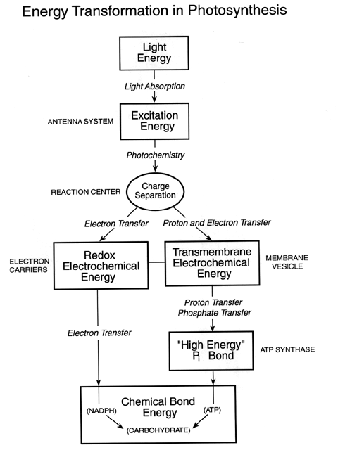
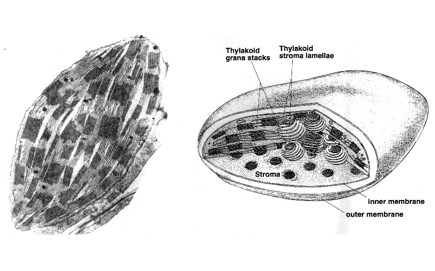
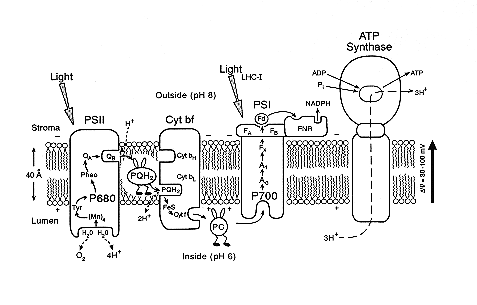
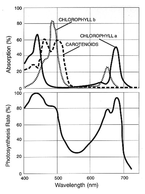
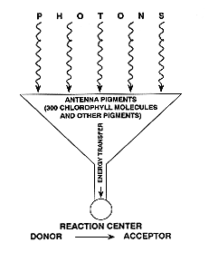
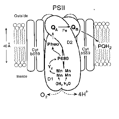
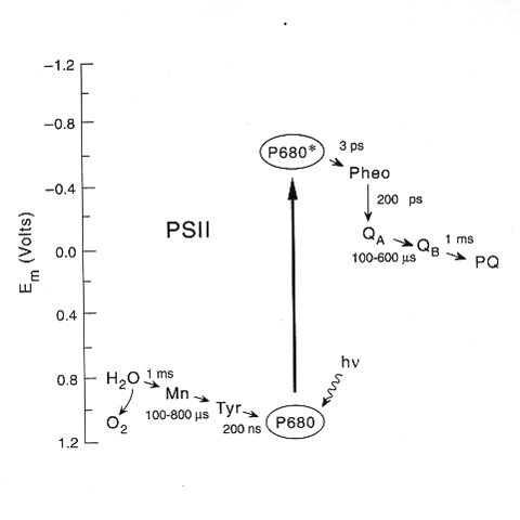
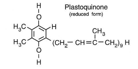
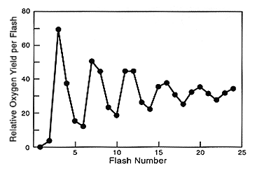
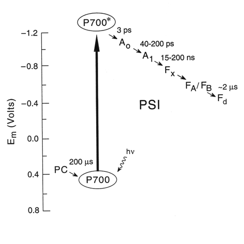
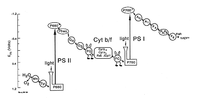
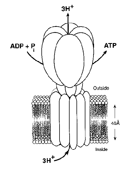
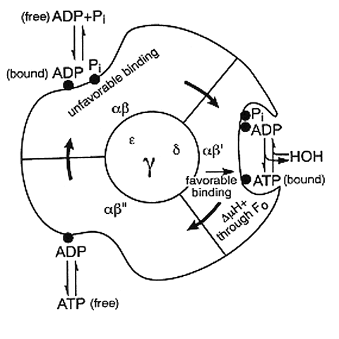
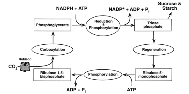
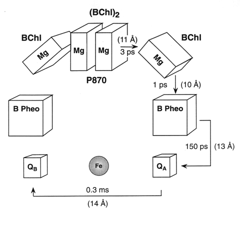
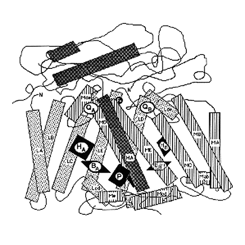
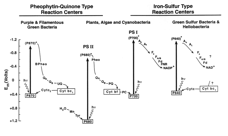